|
BASIC INSTRUCTIONS
To use the basic functionality of GMoS, simply fill the POSITION box with either a single position (e.g. 3.49) or range of consecutive positions (e.g. 3.49-3.51) from a transmembrane segment, using the Ballesteros-Weinstein numbering scheme.
The output will consist in a list of the residues or sequence motifs that appear at this position (or range of positions) in the Class A sequence alignment, ordered according to their abundance. Clicking on the Optionally, the search can be restricted to a specific residue (e.g. D) or sequence motif (e.g. DRX, where X can be used as a wild card), by filling the SEQUENCE MOTIF box.
In this case, the output will consist in a list of the families that present this residue or sequence motif at this position, ordered and colored according to the conservation within the family. Again, clicking on the In either case, the search can be restricted to a specific SUBSET (i.e. the whole Class A alignment or only human sequences without olfactory and orphan receptors) or FAMILY (i.e. amine, peptide, ... receptors) using the pull-down menus. 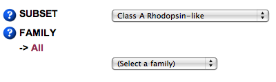
By default, families with no matches in the sequence motif search are not shown in the results. If desired, the whole list of families can be shown by checking the "Show families with 0 matches" checkbox.
For each option, clicking the DETAILED INSTRUCTIONS
The position of the motif in the sequence has to be specified in the Ballesteros-Weinstein numbering scheme [1]. In this numbering method, the position of each residue is described by two numbers: the first (1 to 8) corresponds to the helix in which the residue is located; the second indicates its position relative to the most conserved residue in that helix, arbitrarily assigned to 50. These patterns are easily identifiable on a multiple sequence alignments, and allow an easy comparison among residues in the helical segments of different receptors In bovine rhodopsin, the most conserved residues in each helix are: TM1 Asn55 (1.50)(100%) TM2 Asp83 (2.50) (91%) TM3 Arg135(3.50) (96%) TM4 Trp161(4.50) (88%) TM5 Pro215(5.50) (75%) TM6 Pro267(6.50) (86%) TM7 Pro303(7.50) (97%) hx8 Phe313(8.50) (70%)Thus, for instance, 3.49 corresponds to the residue located immediately before the highly conserved Arg at position 3.50 in the cytoplasmic side of TM3. Obviously, searches are restricted to the positions present in the sequence alignments. Currently, these positions are Complete Class A Human receptors Note that cytoplasmic helix 8 has been also included in the alignment. In this case, the 8.50 position has been assigned to Phe313 (in rhodopsin numbering), as it is present in ~70% of human Class A GPCRs, being the most conserved position in helix 8. Finally, the equivalence between the Ballesteros and other general numbering schemes can be found at this table, created by Elaine C. Meng, at UCSF.
GMoS allows the search of either individual amino acids or sequence motifs of an arbitrary length and complexity. In the latter case, "X" can be used as a wild card. The amino acids must be written in one-letter code, and the field is case-insensitive. For instance, if you are interested in finding conserved sequence motifs starting at the 2.45 position, you could perform the following type of searches: Sequence motif Results
The search can be performed on two different alignments, which can be selected through the SUBSET drop-down box. The default is the "Class A Rhodopsin-like" alignment, retrieved from the GPCRdb, which contains 4998 sequences from several species. Note that this alignment, albeit exhaustive, contains sequence fragments and isoforms plus a considerable amount of redundancy in some families (for instance in chemokine receptors), which can bias the results. To avoid this problems, the search can be performed in an alignment containing only complete and unique human sequences ("Human Class A, no olfactory-orphan"). These sequences have been retrieved from the UniProt database and olfactory or orphan receptors were discarded. The resulting 239 sequences were aligned with ClustalW. Initially, only a small group of proteins were aligned, using a gap-opening penalty of 40.0 and a gap extension penalty of 0.10. Based on this alignment, a mask file for secondary structure was created manually and added to the alignment file. This file was loaded again into ClustalW as a profile, and the remaining sequences were aligned to this profile. As an additional advantage, this alignment results in longer aligned segments, which allows to extend the sequence search near the loops.
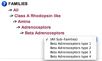
By default, searches are performed in the whole alignment. However, they can be restricted to any specific subfamily (for instance, search only within beta-adrenergic receptors) using the FAMILIES drop-down box. The classification is based on ligand type, according to the families listed in the GPCRdb. Clicking on the "-> All" line will revert the search to the whole alignment. References [1] Ballesteros, J. A.; Weinstein, H. Integrated methods for the construction of three dimensional models and computational probing of structure-function relations in G-protein coupled receptors. Methods in Neurosciences 1995, 25, 366-428. |


|